
84120
Safranin O
suitable for microscopy
동의어(들):
3,7-Diamino-2,8-dimethyl-5-phenylphenazin-5-ium chloride, Safranin, Basic Red 2, Cotton Red, Gossypimine, Safranin T, Safranin Y or A
로그인조직 및 계약 가격 보기
크기 선택
제품정보 (DICE 배송 시 비용 별도)
실험식(Hill 표기법):
C20H19ClN4
CAS 번호:
Molecular Weight:
350.84
색상 지수 번호:
50240
Beilstein:
3924099
EC Number:
MDL number:
UNSPSC 코드:
12171500
PubChem Substance ID:
NACRES:
NA.47
양식
powder or crystals
Quality Level
기술
titration: suitable
pH
10 (20 °C, 10 g/L)
solubility
methanol: 0.01 g/10 mL, red
εmax
≥1000 at 525-535 nm in 50% ethanol
적합성
suitable for microscopy
응용 분야
diagnostic assay manufacturing
hematology
histology
microbiology
저장 온도
room temp
SMILES string
[Cl-].Cc1cc2nc3cc(C)c(N)cc3[n+](-c4ccccc4)c2cc1N
InChI
1S/C20H18N4.ClH/c1-12-8-17-19(10-15(12)21)24(14-6-4-3-5-7-14)20-11-16(22)13(2)9-18(20)23-17;/h3-11H,1-2H3,(H3,21,22);1H
InChI key
OARRHUQTFTUEOS-UHFFFAOYSA-N
유사한 제품을 찾으십니까? 방문 제품 비교 안내
일반 설명
Safranin O, also known as Basic Red 2, is a basic, cationic, and metachromatic azine dye used as a biological stain. The characteristic azine chromophore contains the phenazine, a planar, tricyclic dibenzopyrazine, which consists of two non-planar benzene rings linked by two nitrogen atoms. A delocalized positive charge, an alternation of aromatic and quinonoid structures, and diazotizable amine groups constitute the chromophoric system. c-Methylation makes this dye of moderate overall size. The trimethyl cationic form of safranine is lipophilic, whereas the dimethyl forms are weakly hydrophilic. The dye is commercially available as a chloride.
애플리케이션
Safranin O is widely employed as a microbiological stain, especially as a counterstain in Gram staining to distinguish Gram-positive from Gram-negative micro-organisms, and for staining bacterial spores.
- It is used for human-medical cell diagnosis and serves the purpose of the bacteriological investigation of sample material of human origin.
- It is used to prepare a staining solution, that when used together with other in vitro diagnostic products from our portfolio dyes target structures (e. g. Gram-positive or Gram-negative bacteria, by fixing, where necessary embedding, staining, counterstaining, mounting) in bacteriological specimen materials (for example: smears of bacteriological material that have been air-dried and heat-fixed like sputum, smears from fine needle aspiration biopsies (FNAB), rinses, imprints, effusions, pus, exsudates, liquid and solid cultures), for diagnostic evaluations.
- It is used to demonstrate glycosaminoglycans in human and animal histological specimens and nuclei.
- It is a component of Benda′s polychrome stain.
- It is a component of various polychromes and lignin stains for botanical staining applications.
- It has also been applied for the demonstration of mast cells in cytospin preparations and staining surgical frozen sections.
생화학적/생리학적 작용
In microbiology, Gram staining allows fast differentiation of bacteria as Gram-positive or Gram-negative. The mureine structure of the bacteria cell wall is the basis of the color affinity. In the first step, bacteria will be stained with crystal violet, an aniline dye. After the treatment with iodine solution (Lugol’s solution), a dye-iodine complex will form. During the decolorizing step, this complex stays in the multilayer mureine structure of the cell wall of Gram-positive bacteria and appears blue-violet. Gram-negative bacteria, by contrast, have a cell wall consisting of a single-layered murein structure, and correspondingly re-release the staining dye after treatment with the decoloring solution. Gram-negative bacteria will be counterstained with safranin solution and will then appear pink to red.
특징 및 장점
- Color stable
- Reproducible
- Offers sufficient sensitivity to reveal all structural details
신호어
Danger
유해 및 위험 성명서
예방조치 성명서
Hazard Classifications
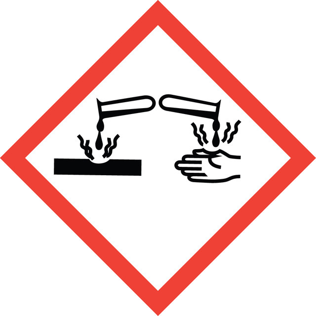
Eye Dam. 1
Storage Class Code
11 - Combustible Solids
WGK
WGK 3
개인 보호 장비
dust mask type N95 (US), Eyeshields, Gloves
Esther Wehrle et al.
Scientific reports, 9(1), 17445-17445 (2019-11-27)
Longitudinal in vivo micro-computed tomography (micro-CT) is of interest to non-invasively capture the healing process of individual animals in preclinical fracture healing studies. However, it is not known whether longitudinal imaging itself has an impact on callus formation and remodeling.
Christian Epple et al.
Biomaterials, 192, 118-127 (2018-11-19)
Large and complex bone defects represent challenging clinical scenarios, typically requiring autologous vascularized bone transplants. In order to bypass the numerous associated limitations, here we aimed at ectopically prefabricating a bone graft surrogate with vascular pedicle. A hollow cylinder of
Finely tuned temperature-controlled cargo release using paraffin-capped mesoporous silica nanoparticles.
Elena Aznar et al.
Angewandte Chemie (International ed. in English), 50(47), 11172-11175 (2011-09-29)
Yee Suan Poo et al.
Journal of virology, 88(12), 6862-6872 (2014-04-04)
Chikungunya virus (CHIKV) is a member of a globally distributed group of arthritogenic alphaviruses that cause weeks to months of debilitating polyarthritis/arthralgia, which is often poorly managed with current treatments. Arthritic disease is usually characterized by high levels of the
José G Raya et al.
Investigative radiology, 46(6), 401-409 (2011-03-24)
To investigate changes of diffusion tensor imaging (DTI) parameters (mean apparent diffusion coefficient [ADC], fractional anisotropy [FA], and first eigenvector) with increasing proteoglycan (PG) extraction of articular cartilage. Twelve cylindrical cartilage-on-bone samples were drilled from 4 human patellae (3 per
자사의 과학자팀은 생명 과학, 재료 과학, 화학 합성, 크로마토그래피, 분석 및 기타 많은 영역을 포함한 모든 과학 분야에 경험이 있습니다..
고객지원팀으로 연락바랍니다.