
S7111
ApopTag Plus In Situ Apoptosis Fluorescein Detection Kit
The ApopTag Plus Fluorescein In Situ Apoptosis Detection Kit detects apoptotic cells in situ by the indirect TUNEL method, utilizing an anti-digoxigenin antibody that is conjugated to a fluorescein reporter molecule.
동의어(들):
Apoptosis detection kit
Quality Level
제조업체/상표
ApopTag
Chemicon®
기술
flow cytometry: suitable
immunocytochemistry: suitable
immunohistochemistry (formalin-fixed, paraffin-embedded sections): suitable
검출 방법
fluorometric
배송 상태
dry ice
일반 설명
The ApopTag Fluorescein In Situ Apoptosis Detection Kit has been tested for specific staining in these model systems: (a) human normal peripheral blood lymphocytes induced with dexamethasone as stained in cytospins, (b) rat regressing mammary gland as stained in formalin-fixed, paraffin-embedded sections, and (c) human leukemic peripheral blood lymphocytes induced with camptothecin, as stained in cell suspensions and used for quantitative flow cytometry.
애플리케이션
ApopTag In Situ Apoptosis Detection Kits label apoptotic cells in research samples by modifying genomic DNA utilizing terminal deoxynucleotidyl transferase (TdT) for detection of positive cells by specific staining. This manual contains information and protocols for the ApopTag Plus Fluorescein In Situ Apoptosis Detection Kit (Catalog number S7111).
Principles of the Procedure
The reagents provided in all ApopTag Kits are designed to label the free 3′OH DNA termini in situ with chemically labeled and unlabeled nucleotides. The nucleotides contained in the Reaction Buffer are enzymatically added to the DNA by terminal deoxynucleotidyl transferase (TdT) (13, 31). TdT catalyzes a template-independent addition of nucleotide triphosphates to the 3′-OH ends of double-stranded or single-stranded DNA. The incorporated nucleotides form an oligomer composed of digoxigenin nucleotide and unlabeled nucleotide in a random sequence. The ratio of labeled to unlabeled nucleotide in ApopTag Kits is optimized to promote anti-digoxigenin antibody binding, or to minimize fluorescein self-quenching. The exact length of the oligomer added has not been measured.
DNA fragments which have been labeled with the digoxigenin-nucleotide are then allowed to bind an anti-digoxigenin antibody that is conjugated to fluorescein (Figure 1A). Fluorescent antibodies provide sensitive detection in immunohistochemistry or immunocytochemistry (i.e. on tissue or cells) and are not subject to experimental variations due to the substrate or the development step. This mixed molecular biological-histochemical systems allows for sensitive and specific staining of very high concentrations of 3′-OH ends that are localized in apoptotic bodies.
The ApopTag system differs significantly from previously described in situ labeling techniques for apoptosis (13, 16, 38, 46), in which avidin binding to cellular biotin can be a source of error. The digoxigenin/anti-digoxigenin system has been found to be equally sensitive to avidin/biotin systems (22). Immunochemically-similar ligands for binding of the anti-digoxigenin antibody are generally insignificant in animal tissues, ensuring low background staining. Affinity purified sheep polyclonal antibody is the specific anti-digoxigenin reagent used in ApopTag Kits and exhibits <1% cross-reactivity with the major vertebrate steroids. In addition, the Fc portion of this antibody has been removed by proteolytic digestion to eliminate any non-specific adsorption to cellular Fc receptors.
제조 메모
2. Protect the anti-digoxigenin fluorescein antibody (#90426) from unnecessary exposure to light.
Precautions
1. The following kit components contain potassium cacodylate (dimethylarsinic acid) as a buffer: Equilibration Buffer (#90416), Reaction Buffer (#90417), and TdT Enzyme (#90418). These components are harmful if swallowed; avoid contact with skin and eyes (wear gloves, glasses) and wash areas of contact immediately.
2. Antibody Conjugates (#90426) and Blocking Solutions (#90425) contain 0.08% sodium azide as a preservative.
3. TdT Enzyme (#90418) contains glycerol and will not freeze at -20°C. For maximum shelf life, do not warm this reagent to room temperature before dispensing.
기타 정보
Reaction Buffer 90417 2.0 mL -15°C to -25°C
TdT Enzyme 90418 0.64 mL -15°C to -25°C
Stop/Wash Buffer 90419 20 mL -15°C to -25°C
Blocking Solution 90425 2.6 mL -15°C to -25°C
Anti-Digoxigenin-Fluorescein* 90426 2.1 mL 2°C to 8°C
Plastic Coverslips 90421 100 ea. Room Temp.
Control Slides 90422 2 ea. Room Temp.
*affinity purified sheep polyclonal antibody
법적 정보
면책조항
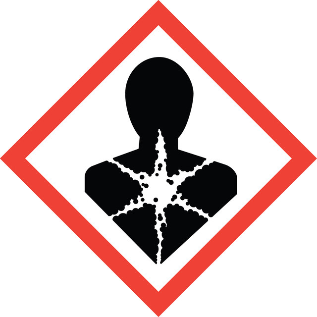
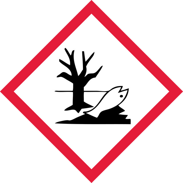
신호어
Danger
유해 및 위험 성명서
Hazard Classifications
Aquatic Chronic 2 - Carc. 1B - STOT RE 2 Inhalation
표적 기관
Respiratory Tract
Storage Class Code
6.1C - Combustible acute toxic Cat.3 / toxic compounds or compounds which causing chronic effects
시험 성적서(COA)
제품의 로트/배치 번호를 입력하여 시험 성적서(COA)을 검색하십시오. 로트 및 배치 번호는 제품 라벨에 있는 ‘로트’ 또는 ‘배치’라는 용어 뒤에서 찾을 수 있습니다.
문서
Cellular apoptosis assays to detect programmed cell death using Annexin V, Caspase and TUNEL DNA fragmentation assays.
관련 콘텐츠
"Recognizing both the tremendous opportunities and the challenges facing cancer research, we are dedicated to developing and refining tools and technologies for the study of cancer. With our comprehensive portfolio, including the Upstate®, Chemicon®, and Calbiochem® brands of reagents and antibodies, researchers can count on dependable, high quality solutions for analyzing all the hallmarks of cancer."
자사의 과학자팀은 생명 과학, 재료 과학, 화학 합성, 크로마토그래피, 분석 및 기타 많은 영역을 포함한 모든 과학 분야에 경험이 있습니다..
고객지원팀으로 연락바랍니다.